news
Publication: Structural and motional changes in a cytochrome c – lipid complex implicated in apoptosis.
Congratulations to lab alum Dr. Mingyue Li and our collaborators on the publication of a new paper in the Journal of Molecular Biology. The paper is online via its DOI link. The paper describes how we used solid-state NMR spectroscopy to characterise the partial destabilization of the native fold of the protein cytochrome c, as it is bound to cardiolipin lipids. This protein-lipid complex is implicated in the process of programmed cell death, where it plays a key role in triggering the self-destruction of undesired or disease cells in higher organisms. Notably, the CL-bound protein catalyses the process of mitochondrial lipid peroxidation, which we see being reconstituted in vitro via mass-spectrometry lipidomics. The latter work is done by our longstanding collaborators in the group of Valerian Kagan at the University of Pittsburgh. The current paper builds on our earlier work, but is of especial interest based on the fact that we bridge some of our prior findings (that suggested the protein to be surprisingly “folded” on the membrane) to studies that report a greater degree of mobility of the CL-bound protein. Here we see how the experimental conditions regulate protein mobility and also pinpoint how different extents of mobility are present in the membrane-bound cytochrome c. Interestingly, the regional dynamics map partly, but not completely onto the previously identified “foldons” that define the folding landscape of this widely studied mitochondrial protein. For more details see the paper below. It is available as #openaccess so just follow the DOI link for access:
Reference:
Mingyue Li, Wanyang Sun, Vladimir A. Tyurin, Maria DeLucia, Jinwoo Ahn, Valerian E. Kagan, Patrick C.A. van der Wel (2021) Activation of Cytochrome C Peroxidase Function Through Coordinated Foldon Loop Dynamics upon Interaction with Anionic Lipids, Journal of Molecular Biology, Volume 433, Issue 15, 23 July 2021, 167057
Publication: New paper on mitochondrial protein-lipid interactions published in PNAS.
Congratulations to lab alumns Dr. Abshishek Mandal and Dr. Jennifer Boatz, as well as our collaborators in the USA and Spain! A new collaborative paper on mitochondrial protein-lipid interactions has just been published in the journal PNAS. In this multidisciplinary work we studied how the protein Drp1 binds the special mitochondrial lipid cardiolipin in order to do its job managing the proper fission of mitochondrial membrane. Our collaborator Rajesh Ramachandran (at Case Western) coordinated a wide array of experimental and computational approaches to determine how Drp1’s “variable domain” (VD) binds cardiolipin. Along the way, two apparent CL-binding motifs were detected, which are seemingly shared by other CL-binding proteins. Their mutation disrupts CL binding and also the proper management of mitochondrial morphology, pointing to the importance of CL-based signals and interactions in this vital cellular process.
Experimentally, our group contributed various solid-state NMR measurements of the lipids, with which we probe the specificity of the interactions and also observe how the protein modulates the lipid bilayer itself. Moreover, we were involved in the sequence/structure analysis: sequence analysis of the “disordered” VD domain pinpointed those parts of the structure most likely to engage in lipid-driven folding upon membrane binding. The “MoRF” motifs (“molecular recognition features”) indeed appear to be involved in CL binding, based on NMR and mutational studies. In Drp1 they are intimately involved in a folding transition of the VD domain upon membrane binding. It will be interesting to see how widespread these CL-binding motifs (CBMs) are in other CL-binding proteins.
Reference:
[1] Mahajan M, Bharambe N, Shang Y, Lu B, Mandal A, Madan Mohan P, et al. NMR identification of a conserved Drp1 cardiolipin-binding motif essential for stress-induced mitochondrial fission. Proc Natl Acad Sci USA2021;118:e2023079118. https://doi.org/10.1073/pnas.2023079118.
Preprint: cytochrome c – cardiolipin studies by ssNMR on bioRxiv
Our latest update on structural studies of this peroxidase-active protein-lipid complex implicated in mitochondrial apoptosis has now posted to bioRxiv: https://www.biorxiv.org/content/10.1101/2021.02.24.432556v1
In the preprint we discuss how ssNMR reveals the involvement of specific and localised dynamics in the lipid-bound protein. Interestingly, the mobility is dependent on the bound lipid species, with an apparent correlation to the resulting peroxidase activity. The lipids thus act as both substrates and regulators of the pro-apoptotic enzymatic activity of the protein. This was also discussed in the recent webinar as discussed in an earlier post.
This work was made possible by several great collaborators at the University of Pittsburgh, and funding from the NIH for both the project and employed instrumentation.
Update: The paper has now been accepted for publication in the Journal of Molecular Biology, and can be found online at its DOI link.
Reference:
Mingyue Li, Wanyang Sun, Vladimir A. Tyurin, Maria DeLucia, Jinwoo Ahn, Valerian E. Kagan, Patrick C.A. van der Wel (2021) Activation of Cytochrome C Peroxidase Function Through Coordinated Foldon Loop Dynamics upon Interaction with Anionic Lipids, Journal of Molecular Biology, in press
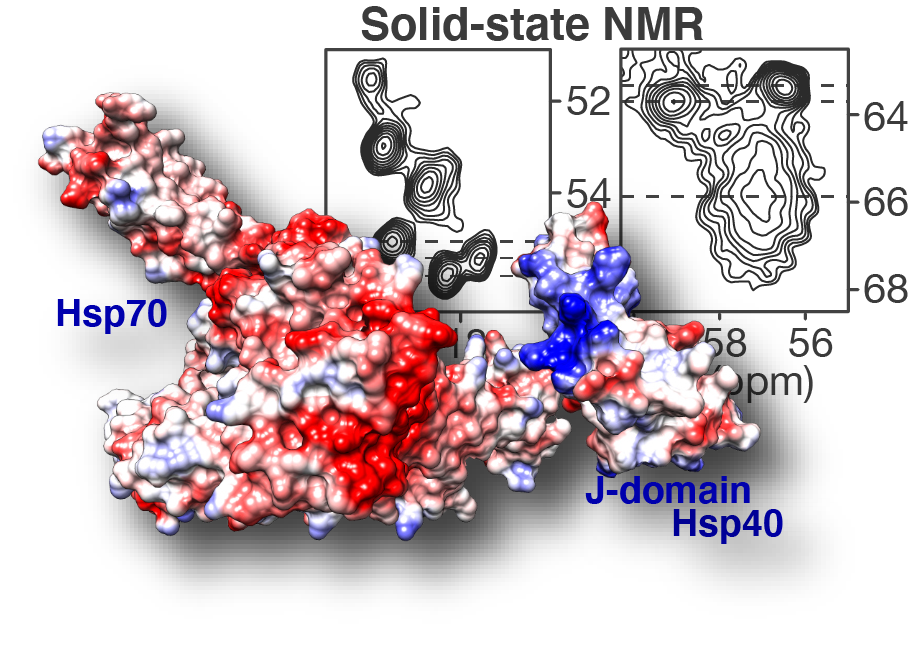
Publication: paper on an anti-polyglutamine oligomeric chaperone studies published!
Congratulations to our postdoc Dr. Irina Matlahov and our collaborators in the group of Prof. Lukasz Joachimiak at UTSW! Our new paper on the structure and function of an oligomer chaperone prohibiting polyglutamine aggregation has now been published in the journal Nature Communications. In it, we deploy a combination of solid-state NMR (ssNMR), solution NMR, cross-linking mass spectrometry (XL-MS) and other methods to visualise and understand these Hsp40 DnaJB8. This so-called co-chaperone teams up with Hsp70 to prevent polyglutamine aggregation in cells, but has been hard to understand. This is because it has itself a strong propensity to self-aggregate (or phase separate), even in a cellular context. Here we deploy a range of methods to identify and characterise an interesting and important feature of this multi domain protein. We show that the protein blocks its own activity when it is just “sitting around”, but can be activated due to environmental triggers. We figure out the specific amino acids are involved, and find that similar interactions are apparently present in other analogous chaperones. The intriguing possibility now arises that this self-blocking allows the chaperones to sit around in a “dormant” state until their action is (urgently?) needed. Then, perhaps, substrate proteins that could cause disease could be bound and that this binding interaction would quickly activate a bunch of these proteins. Further experiments will be needed to test this idea, and whether it can be used to control or activate these innate protective mechanisms as part of disease treatments in the future. Note that polyglutamine proteins aggregate in many incurable diseases, including Huntington’s disease (HD).
For more information, please read our new paper at the journal:
Ryder, B.D.; Matlahov, I.; Bali, S.; Vaquer-Alicea, J.; Van der Wel, P.C.A. & Joachimiak, L.A. (2021) Regulatory inter-domain interactions influence Hsp70 recruitment to the DnaJB8 chaperone. Nat. Commun. 12: 946 [DOI: https://doi.org/10.1038/s41467-021-21147-x]
Our work for this paper was made supported by the American NIH funding our polyglutamine research and instrumentation (grants GM112678 & OD012213-01). Nowadays, our polyglutamine research continues in Groningen with funding from CampagneTeam Huntington and CHDI. Note that this paper is published “open access” like all of our (recent) HD related work.
Note added:
The paper also received some online attention in some science-oriented news sites, based on press releases by the universities involved. This includes a highlight on the HD News website on April 20th.
New (e)book on membrane studies by solid-state NMR released online.
Now available online: we have contributed a chapter to a new (e)book on the topic of solid-state NMR studies of membranes and membrane proteins, edited by Frances Separovic and Marc-Antoine Sani (Univ. Melbourne, Australia). The edited volume “Solid state NMR. Applications in biomembrane structure.” was released in the IOP series in association with the Biophysical Society.
Our chapter (Solid-state NMR studies of peripherally membrane-associated proteins: dealing with dynamics, disorder and dilute conditions [1]) looks at several studies that use ssNMR to probe peripheral membrane proteins. A key focus is on our own work on the mitochondrial protein cytochrome c, and how it binds to cardiolipin lipids during apoptosis, funded by the NIH/NIGMS [2]. In the chapter we try to summarise some of the practical challenges involved, along with potential solutions reported by ourselves and a few other research groups that studied other peripheral membrane proteins by ssNMR.
Cited references:
[1] Van der Wel, P.C.A. (2020) Solid-state NMR studies of peripherally membrane-associated proteins: dealing with dynamics, disorder and dilute conditions. Chapter 10 in Solid-state NMR; applications in biomembrane structure. Edited by F. Separovic & M.-A. Sani; IOP Press (DOI 10.1088/978-0-7503-2532-5ch10)
[2] Li, M.; Mandal, A.; Tyurin, V. A.; DeLucia, M.; Ahn, J.; Kagan, V. E.; van der Wel, P. C. A. (2019) Surface-Binding to Cardiolipin Nanodomains Triggers Cytochrome c Pro-Apoptotic Peroxidase Activity via Localized Dynamics. Structure 2019, 27 (5), 806-815.e4. (DOI 10.1016/j.str.2019.02.007)
New publication on structure of mutant huntingtin protein from Huntington’s Disease
Congratulations to Dr. Jennifer Boatz and other team members for the acceptance and publication of a nice new paper on the structure of the misfolded mutant protein from Huntington’s disease. The paper is online at the Journal of Molecular Biology. Based on an integration of multiple techniques (NMR, EM, and X-ray diffraction), Jennifer assembled a new structural model of the protofilaments that make up the hierarchical fiber architecture of mutant huntingtin exon 1. This is a timely and important step forward in our understanding of the (mis)behaviour of the HD protein, and in particular how it forms pathogenic inclusions and protein aggregates.
In this new paper and prior work we (and others) have seen that the mutant protein is prone to form a collection of different kinds of aggregates, with each their own structure and functional properties. This is of substantial interest from a disease perspective, as these “functional properties” can encompass different degrees of neurotoxic properties. Surprisingly, Jennifer shows in this paper that one contributor to, or trigger of, huntingtin polymorphism is the concentration of the protein. Further studies will have to explore whether or how this finding impacts efforts to replicate cellular behaviour of the protein in vitro, and how it may affect the toxic properties of the aggregated protein states.
Reference:
Protofilament Structure and Supramolecular Polymorphism of Aggregated Mutant Huntingtin Exon 1. Boatz, J.C., Piretra, T., Lasorsa, A., Matlahov, I., Conway, J.F. & Van der Wel, P.C.A. (2020) J. Mol. Biol., 432(16): 4722-4744. [DOI] (Open Access)
Funding & support: The underlying research in this paper was enabled by funding support from the American NIH/NIGMS (grant R01 GM112678) and funding from the CampagneTeam Huntington in the Netherlands. For more information on the disease, see also our HD page and the CTH website. Other support came from the University of Groningen and the University of Pittsburgh.
Note added: Our Institute also highlighted this paper in a nice summary posted on the Zernike Institute website.
New review paper on ssNMR studies of polysaccharide hydrogels
PhD student Mustapha El Hariri El Nokab has put together a nice new review of solid-state NMR studies of polysaccharide hydrogels, which is now out in the journal Carbohydrate Polymers. This review is part of his research project that make use of solid-state NMR (and other tools) to look at functional polysaccharide hydrogels. This new research direction in the lab also constitutes part of our participation in the new Physics of Cancer (www.phycan.nl) initiative of the Zernike Institute for Advanced Materials.
Please find the open-access published paper at the journal Carbohydrate Polymers:
New Publication on Lipid Oxidation
Now online in the journal Free Radical Biology and Medicine: a new review and perspective article by our collaborator Valerian Kagan (Univ. Pittsburgh). The paper (titled “Redox phospholipidomics of enzymatically generated oxygenated phospholipids as specific signals of programmed cell death”) examines the role of controlled lipid oxidation as a source of vital cellular signals. In particular it reviews recent work showing how cardiolipins and phosphatidylethanolamine lipids are oxidised by enzymes, as triggers of apoptosis and ferroptosis. Interestingly, the enzymatically generated oxidised species are distinct from those generated by spontaneous peroxidation, which may be important for the regulatory role of these species.
For more details, read the whole article here:
Kagan et al. (2020) “Redox phospholipidomics of enzymatically generated oxygenated phospholipids as specific signals of programmed cell death” Free Radical Biology and Medicine, Vol. 147, pp. 231-241
Publication: New review article on the structural biology of Huntington’s disease.
Now online at the journal Experimental Biology and Medicine: our new review article summarizing recent contributions from solid-state NMR and electron microscopy to further our understanding of the (mis)behavior of the mutant proteins behind Huntington’s disease.
Citation:
I. Matlahov & P.C.A. van der Wel (2019) Conformational studies of pathogenic expanded polyglutamine protein deposits from Huntington’s disease. Exp. Biol. Med. in press; DOI: 10.1177/1535370219856620
Publication: cytochrome c-cardiolipin complexes as pro-apoptotic lipid peroxidase.
Our first paper of 2019 has just appeared online in the journal Structure. It describes the very nice solid-state NMR studies performed by Dr. Mingyue Li, on a protein-lipid complex involved in the early stages of mitochondrial apoptosis. Together with our collaborators in the groups of Valerian Kagan and Jinwoo Ahn (University of Pittsburgh), she looked at the structure and function of the peroxidase active cytochrome c in its membrane-bound state. For more details on the findings, including how the lipid substrate cardiolipin forms membrane nano domains and acts as a dynamic regulator, please see the paper at the journal. This work was made possible by NIH funding (NIGMS R01 GM113908).
Reference:
Surface-Binding to Cardiolipin Nanodomains Triggers Cytochrome c Pro-apoptotic Peroxidase Activity via Localized Dynamics. Li, M., Mandal, A., Tyurin, V.A., Delucia, M., Ahn, J., Kagan, V.E., & Van der Wel, P.C.A. (2019) Structure, in press [URL]
Additional information/press info: