news
Publication: Impact of aggregation inhibitors on protein aggregation and aggregate toxicity.
Congratulations to Greeshma Jain, Marina Trombetta-Lima and the whole team of co-authors for the publication of our most recent paper on Huntington’s disease (HD), in the journal Nature Communications. This paper is actually the fourteenth peer-reviewed publication from the lab on HD research, since our first paper in 2011. It was enabled by crucial funding from the CampagneTeam Huntington foundation, who supported our HD research program when the Van der Wel group moved back to the Netherlands.
In this new report we describe our use of structural analysis (NMR and EM) to look how aggregation-inhibiting small molecules (polyphenols) change the structure of protein aggregates formed by fragments of the huntingtin protein. We observed that these molecules are effective at slowing down huntingtin exon 1 aggregation, even at sub-stoichiometric concentrations. However, aggregates still formed. Notably, these ‘break-through’ aggregates seem to adopt a different conformation, resulting from a change in the aggregation mechanism. Moreover, we saw that the apparent toxic or pathogenic effects of these aggregates differ from normal aggregates, when we exposed neuronal cells to them. What precisely makes the differ in the reduced toxicity remains a bit unclear, requiring further research.
However, a notable and more general finding is that we need to pay attention to changes in aggregate polymorphs when developing aggregation inhibitors. Here we see a reduced toxic effect, but it seems conceivable that certain inhibitors may redirect the aggregation pathway to more toxic species. Further research would be important for better understanding the interplay of delayed aggregation, changes in aggregate morphology and cellular impacts of protein aggregates.
For more info, please see the actual paper at the journal, with citation below:
Jain G, Trombetta-Lima M, Matlahov I, Ribas HT, Chen T, Parlato R, Portale G, Dolga AM, Van der Wel PCA. Inhibitor-based modulation of huntingtin aggregation mechanisms mitigates fibril-induced cellular stress. Nat Commun. 2025 Apr 15;16(1):3588.
Publication: Structure of protein aggregates implicated in Huntington disease
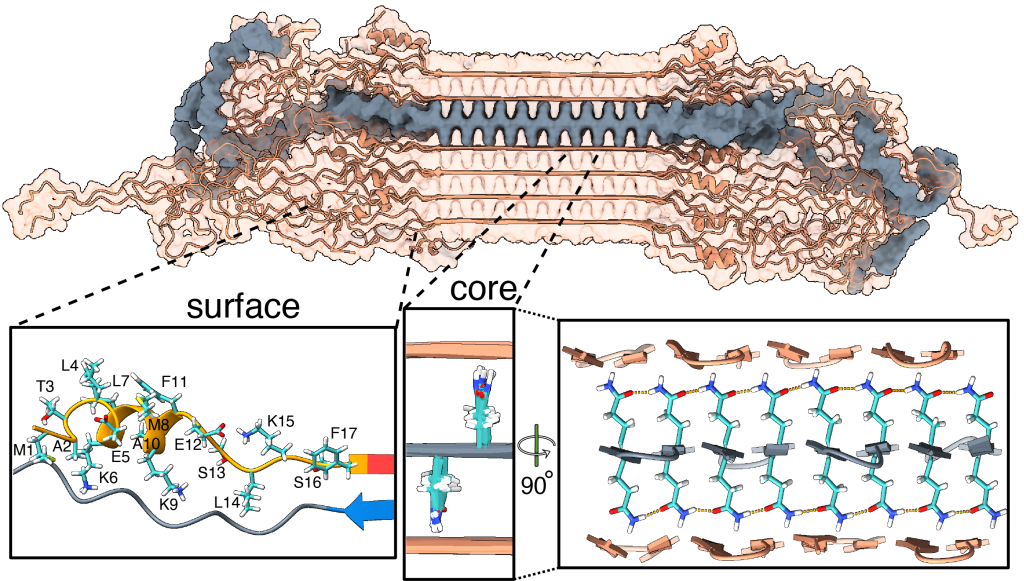
Congratulations to Irina, Greeshma, and Raffaella, together with our international network of collaborators, on a newly published paper that came out in Nature Communications in Dec 2024, in a special collection on the Biology of rare genetic disorders. In this collaborative work, we looked at the structure of proteins behind Huntington’s disease (HD), enabled by crucial funding from the CampagneTeam Huntington and the CHDI Foundation. HD is an inherited neurodegenerative disease, with patients having a mutated form of the huntingtin (HTT) gene. The mutation affects a repeating sequence of CAG codons that has been expanded in HD patients. This translates to huntingtin (HTT) proteins featuring an expanded polyglutamine (or polyQ) segment. Most people have about 20 residues in their HTT polyglutamine segment. Patients have more than 35. Postmortem analysis of HD patient brains reveals the presence of protein clumps (aggregates or inclusions) that contain short fragments of the mutated HTT protein.

In past work we (and others) have studied how HTT fragments also form aggregates when you make the proteins in the lab, and do so more and more aggressively as you expand their polyglutamine segments. Although the precise role of these protein clumps in the disease remains under active debate, there is a consensus that they represent an important biomarker of the disease. Until now, we have lacked a solid understanding of the molecular conformation of the HTT protein in these clumps. For some years now my group (with various collaborators) has been trying to address this question.
Our new paper (together with the team of Markus Miettinen, University of Bergen) introduces the first atomic-level description of the HTT fragments that are increasingly thought to be relevant to the disease. We worked closely together to combine ssNMR data with results from other techniques to build a comprehensive and integrative model that visualizes these protein fibrils in atomic detail. Key aspects of the resulting structure are of course informed and determined by our experimental findings. However, the modeling also unveiled important and useful new insights.
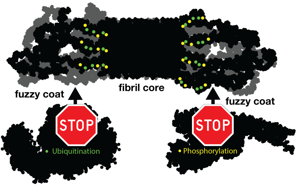
For instance, let’s look at the fuzzy coat that covers these HD amyloid fibrils. Regions outside the mutated polyQ stretch (known as ‘flanking segments’) form a disordered, but densely packed, brush on two sides of a roughly rectangular fibril core. We discuss in the paper how the dense packing reduces or prohibits interactions of other proteins with the flanking segments. This limits the ability of kinases to phosphorylate the aggregated proteins; and similarly for poly-ubiquitination. This may be seen as bad news, as both processes may be used to enable destabilization or destruction of the aggregates.
However, these brushes cover only two sides of the fibril core. The other two sides feature an extensive interface directly between the polyQ amyloid core and the surrounding aqueous phase. In laboratory experiments, it is this type of interface that is targeted by special ‘amyloid dyes’ like thioflavin T (ThT) or Congo red. Previously we had a particularly poor understanding of this part of the fibril surface, mostly because its NMR signals are easily overwhelmed by the much larger core that is sequestered from the surrounding water. The MD shows that this surface is much more dynamic than the fibril core, while it still maintains (on average) some of its structural features (in terms of the glutamine side chain configurations).
To also look at this experimentally, we thought of a way to see the surface through an isotope-editing approach: we can exchange key hydrogens (1H) in the fibril core with deuterium (2H), which makes them invisible in 1H-detected NMR. Then, we could back-exchange the surface with 1H hydrogens, and make the surface visible again, while the core remains invisible. (And vice versa, of course). These NMR measurements were performed in Utrecht, with the team of Markus Weingarth. The great complementarity of the MD and NMR approaches now gives us a new understanding of this part of the fibril surface. Aside from being bound by dyes like ThT, there is an interest in this surface for the design of PET ligands to be used in pre-clinical research, and perhaps ultimately clinical trials. (E.g in HTT lowering drug treatments it would be invaluable to be able to monitor the level of HTT inclusions during treatment)
In any case, we hope that our work can help the HD field in deepening our understanding of the life cycle of HTT and its fragments. Of course, there are also many open questions that remain. One obvious one is that these are proteins that aggregated in laboratory conditions. What happens in a real patient? Would these aggregates be similar or different? We discuss some considerations about this in the paper. Please see the paper for more details, and let us know if you have questions. (The code and coordinates are available on Zenodo)
Citation: Bagherpoor Helabad, M., Matlahov, I., Kumar, R. et al. Integrative determination of atomic structure of mutant huntingtin exon 1 fibrils implicated in Huntington disease. Nat Commun 15, 10793 (2024). https://doi.org/10.1038/s41467-024-55062-8
PDB coordinates of the HTTex1 fibril: download

Publication: NMR of 13C-labeled hyaluronic acid hydrogels (in context of ECM)

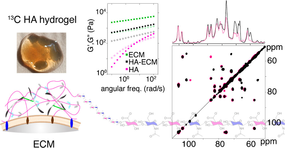
Congratulations to PhD student Pushpa Rampratap (and her co-authors!) on the publication of a new paper on the use of solid-state NMR spectroscopy to study hydrogels that mimic aspects of the extracellular matrix (ECM). Building on her previously published approach to produce 13C labeled hyaluronic acid (HA; with very high molecular weights), she performed extensive series of magic-angle-spinning NMR analyses of HA hydrogels under various conditions. Notably, this included ECM-mimicking conditions that are commonly used in cell culture and biomedical engineering studies (using the Geltrex ECM extract).
The resulting (very nice) paper shows the power of combining 13C enrichment with modern MAS NMR to gain truly atomic-level insights into the behavior of complex hydrogels (or ECMs). Surprisingly (to us), we observed highly localized changes affecting specific atoms in the HA, with the affected atoms being different from what we had expected. We briefly discuss the implications of this finding for e.g. HA-interacting proteins in a biological context. That said, these methods should be particularly powerful also for studying engeneering HA-based hydrogels and nanoparticles, which are finding widespread uses in different types of industries.
Citation:
Rampratap et al. (2024) Resolving Atomic-Level Dynamics and Interactions of High-Molecular-Weight Hyaluronic Acid by Multidimensional Solid-State NMR ACS Appl. Mater. Interfaces 2024, 16, 33, 43317–43328

Funding: new grant from the Barth Syndrome Foundation
We are excited to have received a Development Award grant from the international Barth Syndrome Foundation, enabling us to continue and develop our work studying the molecular mechanisms of this inherited lipid metabolism disease. This is in a sense a follow-up to our recent collaborative publication on this topic. In our paper in Nature Metabolism we used magic-angle-spinning NMR spectroscopy to probe a protein-lipid complex implicated in Barth Syndrome. The NMR analysis showed it to be highly dynamic, with enhanced mobility triggered by the lyso-cardiolipin lipids that are increased in BTHS. Moreover, we showed that it was feasible to probe small molecule interactions with this pathogenic protein-lipid complex, paving the way for further studies.
Some more information on the Foundation’s website. Or see our recent paper for some more background on the science behind the research. Related, earlier, research is also discussed on this page of our website.
Addendum: you can also find a video about the above mentioned paper, hosted by the Barth Syndrome Foundation. In it our co-authors explain and discuss the findings of that work and its relevance to the disease.
Visitor lecture series on solution-state NMR analysis
In the week of May 13-17, we will have an international visitor coming to present a series of educational and research lectures at the University of Groningen. Dr. Andrea Cesari will present a series of four lectures on various aspects of solution-state NMR analysis. Everyone is welcome to attend. For questions please contact the host P.C.A. van der Wel (p.c.a.van.der.wel@rug.nl).
Speaker Dr. Andrea Cesari
Address Fixed-term Junior Researcher (RTDa), Organic Chemist and NMR Spectroscopist at Università di Pisa
Lecture titles:
1. From Sample to Spectrum: good practises in liquid-state NMR Spectroscopy
2. Exploring the NMR toolkit for molecular recognition studies – from relaxation times to 2D maps
3. Applications of liquid-state NMR spectroscopy in pharmaceutical technology
4. Nanoparticle-based chemosensors using NMR spectroscopy
More info here
Publication: New paper about photochemical approaches to studying polyQ protein aggregation.
Congratulations to PhD student Raffaella Parlato, Dr. Jana Volaric, and their collaborators, on the publication of a nice new paper in the Journal of the American Chemical Society. This is the final result of an idea from some years ago, which came together very nicely thanks to a great team of collaborators. The goal of this work was to explore the idea of putting polyglutamine aggregation under some degree of photo-control. In prior work, it has been shown that b-hairpin formation is a key step in the aggregation process of expanded polyQ proteins. Azobenzene-based groups can be used to favor or disfavor beta turn structures under the influence of light, but prior ways of implementing this have been limited in various ways. A new amino acid analogue is introduced with more favorable structural and photochemical properties. And, we tested it in the context of polyQ peptides, seeing it indeed modulating the conformation of resulting aggregates (seen by ssNMR on unlabeled peptide fibrils!). For more details, see the paper. It is open access, so easily accessible.
Citation:
Raffaella Parlato, Jana Volarić, Alessia Lasorsa, Mahdi Bagherpoor Helabad, Piermichele Kobauri, Greeshma Jain, Markus S. Miettinen, Ben L. Feringa*, Wiktor Szymanski*, and Patrick C. A. van der Wel* (2024) J. Am. Chem. Soc. 2024, 146, 3, 2062–2071. Photocontrol of the β-Hairpin Polypeptide Structure through an Optimized Azobenzene-Based Amino Acid Analogue. DOI: https://doi.org/10.1021/jacs.3c11155
Publication: Review article on polyglutamine protein analysis by solid-state NMR spectroscopy.
Our new review article summarizing progress in polyglutamine protein studies by solid-state NMR is now available on the website of the journal Biochemical Society Transactions. This is an invited mini-review that was requested to cover the progress made in our understanding the aggregation and misfolding of mutant polyglutamine proteins implicated in diseases such as Huntington’s disease and several versions of spinocerebellar ataxia (SCA). We describe how studies from ssNMR has been used to better understand the structure of these protein aggregates, and thus helps us analyze how the aggregation process occurs and how aggregates may play a role in these diseases. The paper is published open-access, so you should be able to read it to see all the details. (Naturally, the format of the mini-review limited the word count and meant that much had to be omitted)
Citation:
Patrick C.A. van der Wel; Solid-state nuclear magnetic resonance in the structural study of polyglutamine aggregation. Biochem Soc Trans 2024; BST20230731. doi: https://doi.org/10.1042/BST20230731
Publication: collaborative paper on perovskite-related materials, with Sn and Se ssNMR
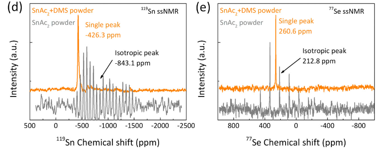
Congrats to our collaborators from the group of Maria Loi and others for the publication of their new paper on in situ SnSe deposition as passivation for scalable and stable quasi-2D lead–tin perovskite solar cells, in the journal Energy & Environmental Science. Dr. Lasorsa in our group contributed tin and selenium ssNMR analysis to this interdisciplinary paper. For more information see the paper at the journal.
Publication:
Chen L, Tekelenburg EK, Gahlot K, Pitaro M, Xi J, Lasorsa A, et al. In situ SnSe deposition as passivation for scalable and stable quasi-2D lead–tin perovskite solar cells. Energy Environ Sci. 2023;10.1039.D3EE02507A.

Upcoming Symposium Dutch Protein Aggregation Network DPAN
Hold the date! On November 30th there will be an inaugural symposium of the Dutch Protein Aggregation Network (DPAN), sponsored by NWO. This one-day even (free registration) will be held at the VU in Amsterdam. It welcomes all Dutch researchers interested in protein aggregation related to human disease (including PIs, PhD students, postdocs..).
The DPAN network aims to bring together the protein aggregation researchers of The Netherlands. With the help of NWO sponsorship DPAN will start with a one-day symposium on Nov 30th. This will take place at the O|2 building of the VU in Amsterdam.
The keynote speaker will be Harm Kampinga from the UMCG Groningen. Many other speakers will discuss their research on amyloid formation and other aspects of protein aggregation.
Please join us and register here.