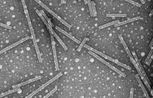
The determination of protein structures in three-dimensional, molecular detail is critical to our understanding of proteins’ normal function and their role in disease. Solid state magic angle spinning (MAS) NMR plays an important role in permitting the determination of protein structures under sample conditions that are otherwise inaccessible to atomic-resolution characterization. In particular, it allows the study of immobilized or aggregated proteins, for instance as amyloid-like fibrils. The study of amyloid fibril structure and formation is of great interest, due to its medical significance (in diseases such as Huntington’s and Alzheimer’s disease) but also for its contribution to our understanding of protein folding and misfolding in general.

Huntington’s disease (HD) is associated with the mutation and aggregation of the huntingtin protein, by expansion of a naturally occurring polyglutamine (polyQ) segment. Unfortunately, the exact mechanism of aggregation, the fibrillar structure as well as the primary source of toxicity all remain enigmatic, both for HD and for amyloid diseases in general. Through the use of MAS ssNMR we investigate the structural and dynamic features of amyloid fibrils, in order to elucidate features of amyloid structure and formation. These investigations have provided direct insight into the structure of the mature fibrils and the impacts of the non-polyQ flanking domains found within huntingin’s exon-1 segment [1-4].
Amyloid formation appears to be common to a range of different proteins and peptides, which we have explored in earlier work [5,6]. Our amyloid studies are accompanied by ongoing improvements in sample preparation and spectroscopic techniques. It is important to note that not all protein aggregation processes result in amyloid formation. Commonly, amorphous looking aggregates are formed, for instance by proteins associated with congenital cataract formation. Despite their appearance, such aggregates can retain a well-defined homogeneous internal structure on the atomic level [7].
Related publications
- Sivanandam et al. (2011) The Aggregation-Enhancing Huntingtin N-terminus is Helical in Amyloid Fibrils. J. Am. Chem. Soc. 133(12): 4558–4566
- Kar et al. (2013) β-hairpin-mediated nucleation of polyglutamine amyloid formation. J. Mol. Biol., 425(7):1-45
- Mishra et al. (2012) Serine phosphorylation suppresses huntingtin amyloid accumulation by altering protein aggregation properties. J. Mol. Biol., 424(1-2), p. 1-14
- Hoop et al. (2014) Polyglutamine amyloid core boundaries and flanking domain dynamics in huntingtin fragment fibrils determined by solid-state NMR. Biochemistry 53(42): 6653-6666
- Li et al. (2011) Amyloid-like fibrils from a domain-swapping protein feature a parallel, in-register conformation without native-like interactions. J. Biol. Chem. 286(33); 28988-28995 [at journal][PubMed]
- Van der Wel, P.C.A. (2012) Domain swapping and amyloid fibril conformation. Prion: 6(3): 211-216.
- Boatz, J.C., Whitley, M.J., Li, M., Gronenborn, A.M., & Van der Wel, P.C.A.# (2017) Cataract-associated P23T γD-crystallin retains a native-like fold in amorphous-looking aggregates formed at physiological pH. Nat. Commun. 8:15137. (DOI)